The Role of Eye Pressure Testing in Early Detection of Open-Angle Glaucoma
The Importance of Eye Pressure Testing for Early Glaucoma Detection
Eye pressure testing is a quick, non-invasive way to spot the warning signs of open-angle glaucoma—the most common form of glaucoma. Often symptom-free until vision loss occurs, open-angle glaucoma can progress silently, making early testing essential to prevent irreversible damage.
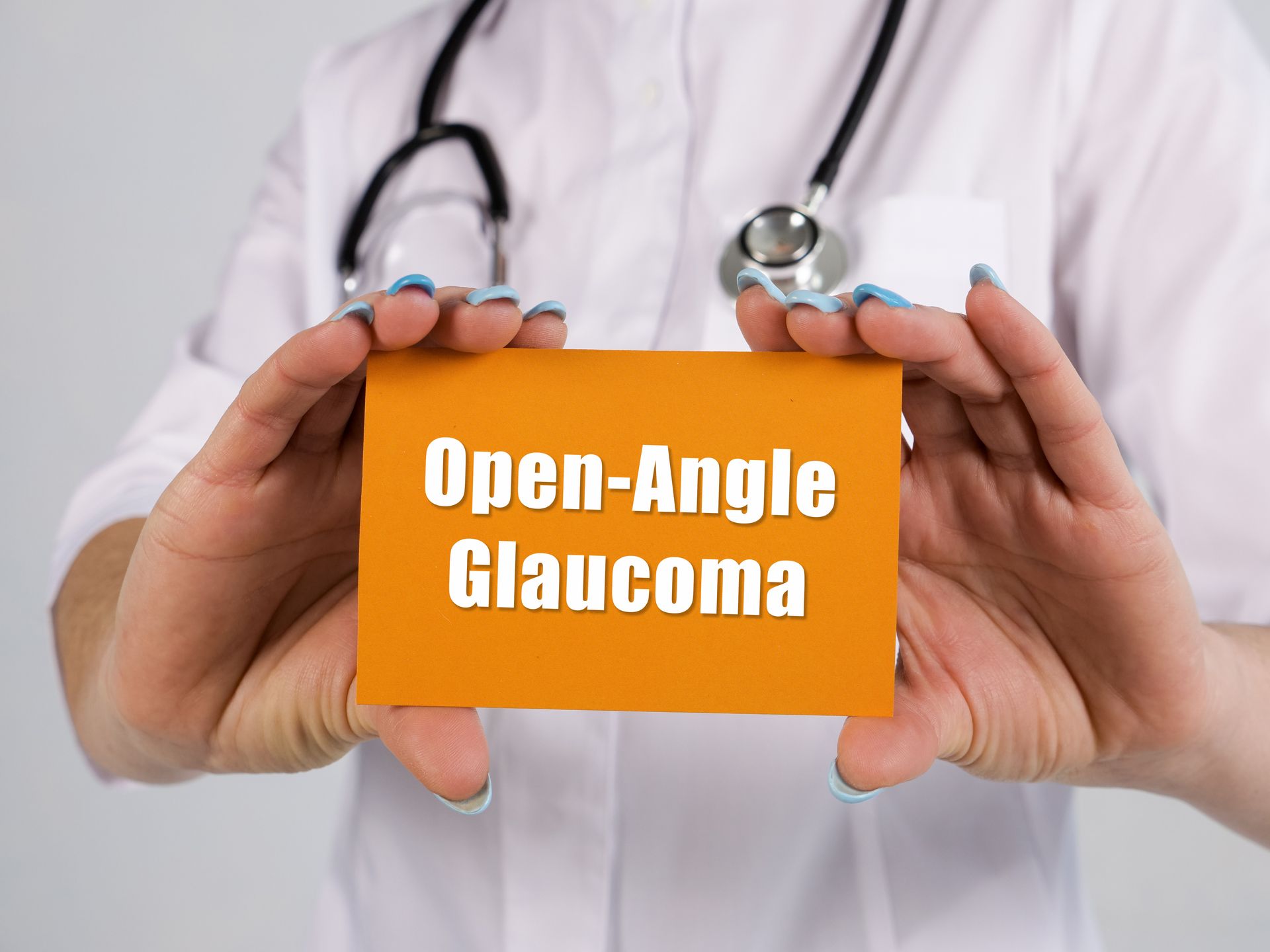
Understanding Open-Angle Glaucoma
Open-angle glaucoma occurs when the drainage canals in the eye become partially blocked, causing fluid to build up and increase intraocular pressure. While anyone can develop this condition, certain risk factors make some individuals more susceptible.
These include age (especially those over 40), a family history of glaucoma, and certain ethnic backgrounds, including African, Hispanic, or Asian descent.
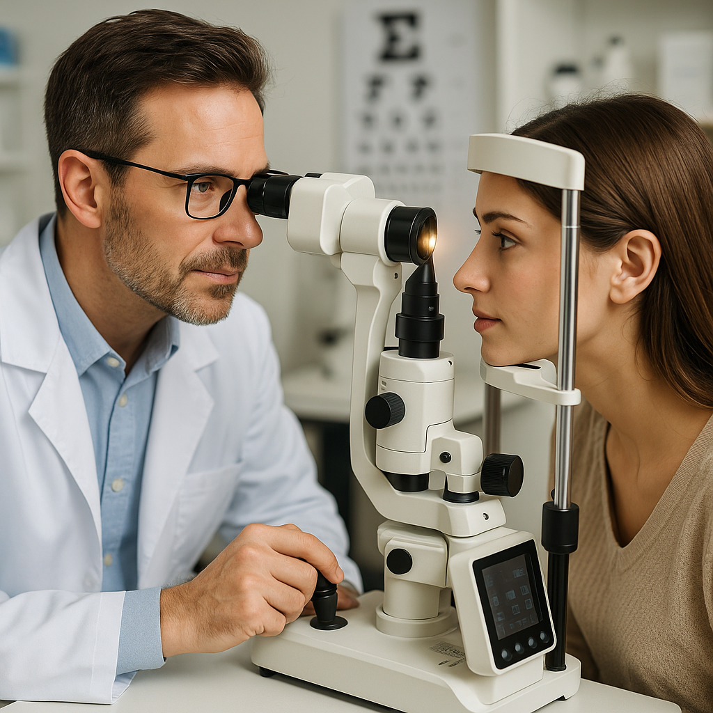
The Role of Eye Pressure Testing (Tonometry)
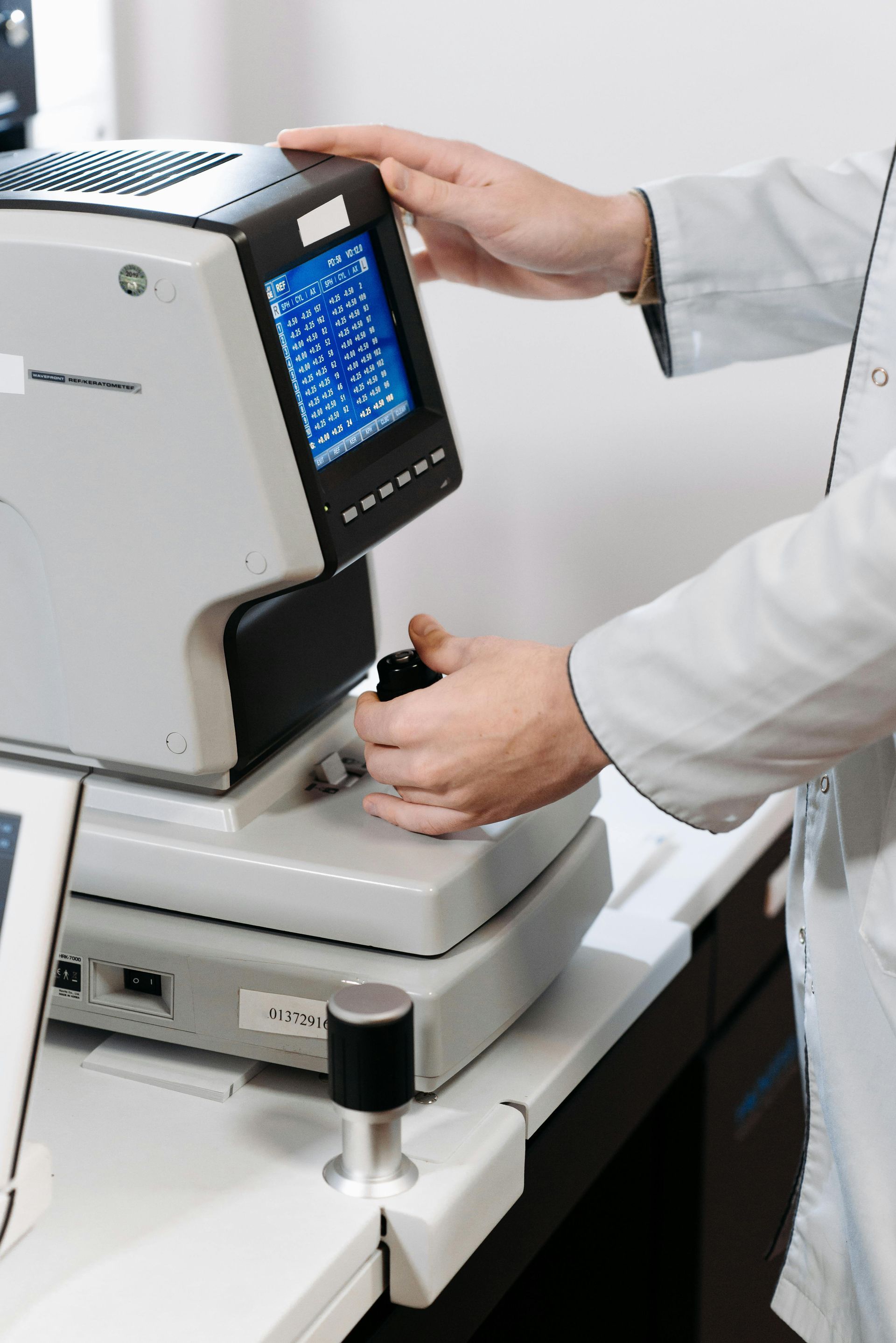
Tonometry is an important tool in assessing intraocular pressure and detecting early glaucoma symptoms. Recent developments in tonometry and eye imaging have made the test more accurate and less invasive. When paired with additional eye tests, this pressure test greatly improves the likelihood of identifying open-angle glaucoma in its earliest stages.
Establishing Baseline Measurements
When you first undergo the test, optometrists establish a baseline IOP reading. This initial measurement is important for subsequent evaluations, as it helps healthcare professionals monitor changes in pressure over time.
An individual's normal range is between 11 and 21 mm Hg, which can alert the patient and doctor to potential issues. If the baseline IOP is high, additional testing may be required to assess the risk of developing glaucoma.
Comprehensive Eye Exams for Health Monitoring
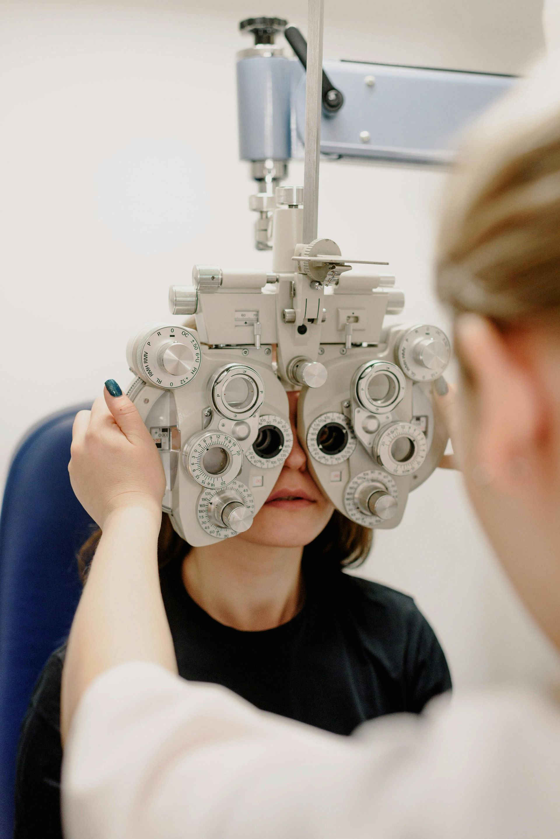
Incorporating eye pressure testing into a comprehensive eye exam allows for a thorough view of eye health. Along with tonometry, your optometrists may perform visual field tests and retinal examinations to assess overall eye function. Through this multifaceted approach, any changes in eye pressure are explained within the patient's complete eye health profile.
Advances in Technology and Techniques
Devices like optical coherence tomography (OCT) provide detailed imaging of the optic nerve and surrounding tissues, complementing traditional tonometry. These innovations allow doctors to detect subtle changes that might indicate the early stages of open-angle glaucoma, improving early intervention strategies and improving your eyesight.
Treatment Options Following High IOP
The primary goal of tonometry is to lower intraocular pressure to prevent damage to the optic nerve. Treatment may include prescription eye drops, which help reduce fluid production or improve drainage. In some cases, oral medications or laser treatments may be appropriate. Close monitoring of IOP after initiating treatment is essential to assess its effectiveness and make necessary adjustments.
Monitoring Changes and Optic Nerve Health
Regular eye pressure testing allows specialists to track fluctuations in IOP. For instance, even if you initially present with normal pressure, routine testing can reveal subtle increases. These changes can indicate the early stages of open-angle glaucoma, even before any noticeable symptoms arise.
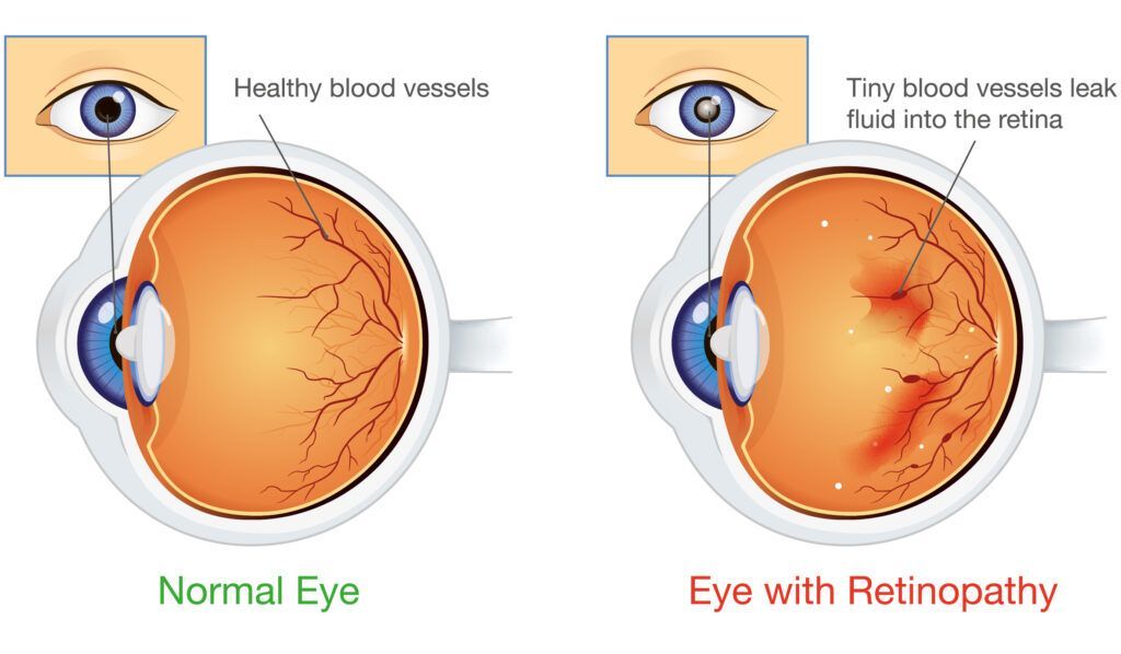
In addition to measuring IOP, tonometry often coincides with examinations of the optic nerve. The health of this nerve is directly impacted by intraocular pressure, which when increased can lead to compression and damage. Your optometrist may use a combination of tonometry and detailed imaging of the optic nerve to evaluate its condition.
If you haven’t had an eye pressure test recently, consider scheduling one soon with Optometric Associates of Southern Maine. Our team of experts assesses for open-angle glaucoma risk to prevent irreparable damage to your eye. Schedule your test